Division of Infectious Diseases

Baloxavir Marboxil for Uncomplicated Influenza – Worth the Cost?

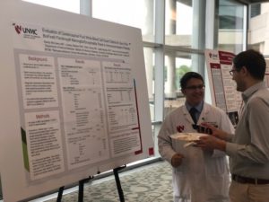
Influenza season is in full swing and with that, the discussions surrounding treatment are heating up! Dr. Hankins, a second year ID fellow, led our recent journal club discussion on Baloxavir.

The New England Journal of Medicine article, Baloxavir Marboxil for Uncomplicated Influenza in Adults and Adolescents, discusses two randomized control trials, that were double-blinded for healthy outpatients with acute, uncomplicated influenza during the 2016-2017 influenza season. Phase II & Phase III trials reported. The primary outcome of both trials was time to alleviation of symptoms, with secondary outcomes of viral RNA titers, duration of virus detection, and resistance testing. The Phase II trial evaluated one time doses of baloxavir 10mg, 20mg and 40mg, vs placebo. The study evaluated adults in Japan who had a fever, at least one respiratory symptom, one systemic influenza symptom, and a positive rapid antigen test (the standard of care in Japan). The results of the phase II trial showed baloxavir significantly reduced time to alleviation of symptoms, with 54.2 hours in the 10 mg group, 51.0 hours in the 20 mg group, and 49.5 hours in the 40 mg group, compared to 77.7 hours in the placebo group.

The phase III trial evaluated baloxavir vs oseltamivir vs placebo, and used similar inclusion criteria, although it did not require a positive influenza antigen test, as this study was in the United States as well as Japan. It evaluated a one time dose of baloxavir 40mg or 80mg (decided by weight) vs oseltamivir 75mg BID for 5 days vs placebo. In the phase III Baloxavir successfully decreased time to alleviation of symptoms of approximately 1 day compared to placebo, but the time to alleviation of symptoms was comparable to oseltamivir. Viral load reduction appeared to be greater in the first few days, but then becomes equitable with oseltamivir. Plus, resistance to Baloxavir may increase to 10% after a single dose. Adverse events were similar in all groups.

Take-always: Baloxavir appears similar to oseltamivir for treatment of outpatient symptoms of influenza. Is there a possible utility for the viral load reduction of baloxavir in the inpatient setting, particularly among the immunocompromised or critically ill? Is the one-time dose of Baloxavir worth the cost given the no change in symptoms for outpatients? If we use it, will it only be short-lived given the higher rate of resistance that seems to evolve?

Will we prescribe it? The overall consensus seemed to be no; not if we can still use oseltamivir.


It’s Getting Hot in Here: The Conundrum of Fever in the ICU

Fever has plagued mankind through the ages although was not until the 1600s when Thomas Sydenham reportedly first recognized that fever was an innate response” to get rid of the injurious agents causing the disease”.

In the intensive care unit, fever is one of the most common abnormal signs documented and frequently results in changes in clinical management of the patient, yet we continue to lack adequate evidence regarding the definitions of fever, the incidence, the impact of fever on mortality and whether or not we should treat fever (and if we should, in who).

With this in mind, clinicians need to review fever and the continual evolution of literature regarding this, particularly as it is very likely that temperature management will continue to garner attention regarding how temperature impacts sepsis management and outcomes.

Definition and Pathophysiology

Normothermia: Defined as a normal body temperature of approximately 37.0 C although there is noted variation that is normal of+/- 0.5C throughout the day.

  • Fever: There has been a range of temperatures defined as fever in the literature, however a core body temperature of > 38.3 C has been accepted as fever among the critically ill. However, it should be noted that lower temperatures may represent fever in the immunocompromised.
  • High Fever: Generally this is defined as a persistent body temperature greater than 39-40 C, although here again there has been some variations in the literature.
  •  Prolonged Fever: If the duration of fever is >5 days, it is often considered a prolonged fever.
  • Hyperthermia: An elevated body temperature, often >41 C,  that may be indistinguishable from “fever” clinically, but is due to lack of change in the hypothalamic “setpoint” (See below for further detail).

Pathophysiology

Fever and hyperthermia are notably different mechanisms of alterations in body temperature, although initially these may be difficult to differentiate during a patient evaluation. Thermoregulation is controlled by the hypothalamus, and in fever from infectious and noninfectious causes, the “set point” of temperature by the hypothalamus is increased thereby “allowing” fever. This however is not the case in hyperthermia syndromes. In hyperthermia, there is no change in the hypothalamic “set point” and body temperature is dysregulated resulting in decreased ability for natural heat dissipation.

Measurement of Fever

With the definition of fever behind us, we can focus on how exactly we detect fever. The gold standard for measuring temperature is through a pulmonary artery catheter. However, in the absence of this, we resort to alternative measurements such as thermistors on bladder catheters, esophageal probes or rectal probes. Alternative options also include tympanic membrane or temporal artery thermometers. Of these, rectal, esophageal, bladder and tympanic membrane (not infrared) may be the closest to core body temperature. Axillary, oral and skin temperatures are available but may be impractical and/or less reliable.

Incidence of Fever

Body temperature measurements are ubiquitous in the hospital and intensive care units. Fever is also incredibly common with reports of approximately 25 to greater than 80% of ICU admissions having at least one fever during their ICU stay, and of this approximately 50% are ultimately attributed to infection.

Causes of Fever: Infectious vs Noninfectious vs Hyperthermia

There are many potential causes of fever in the ICU. When evaluating patients, history can be critical to determining if there is a risk for a hyperthermia syndrome such as heat stroke, malignant hyperthermia, neuroleptic malignant syndrome or serotonin syndrome or endocrine diseases causing hyperthermia such as drug intoxication and withdrawal,thyrotoxicosis, adrenal crisis and pheochromocytoma.

Infectious causes a fever are incredibly diverse and may be secondary to a variety of pathogens including bacteria, viruses, protozoa and fungi. Further, some of these fevers will be driven by infections that were present are incubating on arrival to the ICU and others will develop during the ICU stay as a nosocomial infection.

Finally, there are also many noninfectious causes of fever among critically ill. These can include post-operative fevers, drug reactions, thrombosis and emboli or infarct, hemorrhage, pancreatitis, transfusion reactions, pancreatitis or acalculus cholecystitis and occult malignancy among many other potentials.

Given the robust array of potential causes of fever or hypothermia in the critically ill population, providers need to be very thoughtful and thorough when obtaining the initial history, past medical history, review of systems, medications, social and exposures/travel history combined with a thorough examination of the patient to search for potential clues for the cause of fever. Guidelines call for thoughtful evaluation and not to proceed with a reflex fever evaluation.

Managing Fever

Finding fevers, or other abnormal temperatures, among critically ill patient’s is clearly common and impacts clinical decision making however we still lack evidence on whether or not we should intervene on all fevers. The difficulty remains the complex balance between risks and benefits that fever portends to the patient.

The risks and benefit of fever are complex at best. There is always concern for patient discomfort with fever the combined with increased metabolic and oxygen demands raising increased risk of brain damage and multi organ failure. On the other hand, fever is a normal and adaptive response to infection that prompts further evaluation and management from clinicians. Further, this normal febrile response to infection may improve host immune responses, impede bacterial growth, enhance both antimicrobial concentrations and efficacy.

There is ongoing discrepancies in the literature regarding whether or not fever carries increased mortality or not and these variations may be due to timing of fever, severity of fever and underlying comorbidities at the time of fever. In a 2017 meta-analysis, Rombus et al aimed to assess the association between body temperature and mortality among patients with sepsis(10). In this particular study, fever(greater than 38 C) was associated with decreased mortality while hypothermia (<36 C) had a higher associated mortality compared to normothermia. This is concordant with a 2017 study in Critical Care Medicine regarding the predictive value of fever in the emergency department for patients, which demonstrated an inverse relationship between decreased mortality and shorter hospital length of stay associated with higher temperatures on presentation. However, patients with high and prolonged fever have been shown to have higher mortality in some studies.

Treatment of fever in patients with neurologic injury, myocardial ischemia and arguably in severe hypoxia, may improve outcomes, but this does not necessarily translate to all critically ill populations. In addition, hyperthermia syndromes do require emergent therapy which may include stopping the offending agent, antidote and supportive cares including physical cooling. In hyperthermia, antipyretics are not routinely recommended as they are not usually effective in states without an elevated hypothalamic setpoint.

Use of Antipyretics or cooling therapies

Finding fever is one thing and deciding to treat it in order to decrease it is another. This area remains one of “hot” debate including whether fever is simply a marker for poorer outcomes or whether active treatment of abnormal body temperature can improve outcomes.

Given the frequency of acetaminophen administration among critically ill patients with fever, a randomized controlled trial assessed treatment versus placebo and did not find a significant difference in the number of ICU free days or 90 day mortality(12). Systematic reviewed meta-analysis of antipyretic therapy among critically ill septic patients also did not demonstrate a significant improvement in 28 day or hospital mortality(6). However, on the contrary, there has been associations of fever with increased mortality prompting further observational study on the association of body temperature and antipyretics treatments with mortality(13). In this study it was noted that in patients without sepsis, high fever (>39.5) was associated with mortality and antipyretics among septic patients increased mortality, raising question into the potential different impacts fever and antipyretics may have depending on whether the patient has sepsis(13). A recent systematic review and meta-analysis of antipyretic use in critically ill adults with sepsis did not find a significant improvement in either 28-day or hospital mortality(6).

Physical cooling therapies are also utilized in both fever and hyperthermia. There is a vast array of technology from the simplicity of ice packs and stands to advanced cooling blankets to intravascular cooling devices. It is unknown if there is a significant difference with these varying methods for cooling patient’s and despite the increased speed and stability of intravascular cooling, we lack substantial evidence to state that this is provides significant improvement in patient outcome(4, 5). All cooling methods carry some risk however the intravascular devices are by nature invasive in carry the risk of central line placement with them(4).

Summary

What to do with abnormal body temperatures, particularly fever, remain an area where we need more research to guide our clinical practice. For now, abnormal temperatures should be evaluated for the underlying etiology and then the art of medicine kicks in – which temperature is right for your patient? Hot, cold or somewhere in between? The many risks and benefits will need to be weighed for each individual patient – unless you have high fever with a hyperthermia syndrome, neurologic injury, myocardial ischemia or severe hypoxia – in these settings, normothermia may prevent further organ damage.

Content by Dr. Kelly Cawcutt. 


  1. Bryan CS. Fever, famine, and war: William Osler as an infectious diseases specialist. Clinical infectious diseases. 1996;23(5):1139-49.
  2. Laupland KB, Shahpori R, Kirkpatrick AW, Ross T, Gregson DB, Stelfox HT. Occurrence and outcome of fever in critically ill adults. Critical care medicine. 2008;36(5):1531-5.
  3. Laupland KB. Fever in the critically ill medical patient. Critical care medicine. 2009;37(7):S273-S8.
  4. Golding R, Taylor D, Gardner H, Wilkinson JN. Targeted temperature management in intensive care–Do we let nature take its course? Journal of the Intensive Care Society. 2016;17(2):154-9.
  5. Kushimoto S, Yamanouchi S, Endo T, Sato T, Nomura R, Fujita M, et al. Body temperature abnormalities in non-neurological critically ill patients: a review of the literature. Journal of Intensive Care. 2014;2(1):14.
  6. Drewry AM, Ablordeppey EA, Murray ET, Stoll CR, Izadi SR, Dalton CM, et al. Antipyretic Therapy in Critically Ill Septic Patients: A Systematic Review and Meta-Analysis. Critical care medicine. 2017;45(5):806.
  7. Rehman T. Persistent fever in the ICU. CHEST Journal. 2014;145(1):158-65.
  8. O’Grady NP, Barie PS, Bartlett JG, Bleck T, Carroll K, Kalil AC, et al. Guidelines for evaluation of new fever in critically ill adult patients: 2008 update from the American College of Critical Care Medicine and the Infectious Diseases Society of America. Critical care medicine. 2008;36(4):1330-49.
  9. Niven DJ, Laupland KB. Pyrexia: aetiology in the ICU. Critical Care. 2016;20(1):247.
  10. Rumbus Z, Matics R, Hegyi P, Zsiboras C, Szabo I, Illes A, et al. Fever Is Associated with Reduced, Hypothermia with Increased Mortality in Septic Patients: A Meta-Analysis of Clinical Trials. PloS one. 2017;12(1):e0170152.
  11. Sundén-Cullberg J, Rylance R, Svefors J, Norrby-Teglund A, Björk J, Inghammar M. Fever in the Emergency Department Predicts Survival of Patients With Severe Sepsis and Septic Shock Admitted to the ICU. Critical care medicine. 2017;45(4):591-9.
  12. Patel J. Acetaminophen for Fever in Critically Ill Patients with Suspected Infection. The New England journal of medicine. 2016;374(13):1291.
  13. Lee BH, Inui D, Suh GY, Kim JY, Kwon JY, Park J, et al. Association of body temperature and antipyretic treatments with mortality of critically ill patients with and without sepsis: multi-centered prospective observational study. Critical care. 2012;16(1):R33.

Global Burden of Tuberculosis: Are We Making Any Improvements?

UNMC is a proud to play a critical role in biopreparedness and global health, which also means we have to stay up-to-date on global health, including tuberculosis.
In a recent Infectious Diseases journal club, Dr. Lawler presented the following 2018 Lancet article on the global burden of TB.
Take-Home Points:
We still see over 10.4 incident cases of TB globally (HIV and non-HIV patients) with over 1.4 million deaths in 2016. TB remains a critical need and epidemic globally that demands attention.
The vast majority of non-HIV related TB deaths are in those < 65. This relates to significant socioeconomic burdens of disease.
The incidence is decreasing by 1.3% among HIV negative individuals and 4% among HIV positive patients, far below the the goal of 10% decrease by 2030 in the Sustainable Development Goal.
Much of this data is done via modeling, but truth be told, many of model are based on assumptions in areas of the world with high burden of disease and very poor diagnostics for confirming TB. Per Dr. Lawler’s assessment, only approximately 400,000 data points are hard data vs theoretical data with the subsequent modeling. Yet, this is the data driving our guideline development. Thus raising critical, ethical questions of: What if we used the millions of dollars funding this study to provide those diagnostics? Where is the greatest impact for generating support for improving care and reduction of TB?
The study is perhaps a cautionary tale of the dangers of taking modeling data as a hard, cold truth. The highlight – the reminder that TB is still a major player in global disease and death.


Decisive De-labelling in Cancer Patients: Just what the Doctor Ordered

The following was previously posted by Dr. Marcelin to SHEA Journal Club published online in January 2019.

Although 10% of Americans report penicillin allergies, 90% of those allergies are not substantiated. Up to 25% of patients living with cancer report penicillin allergies, but more than half of these are low risk and could tolerate beta-lactams.

Cancer patients are likely to receive inappropriate antibiotics for a documented penicillin allergy because they are immunocompromised, therefore “special”. Additionally, admission for systemic illness like neutropenic fever, it may not be convenient to schedule penicillin skin testing, and other medications could interfere with skin testing.

Trubiano et al. focused specifically on cancer patients to test the feasibility of implementing an oral penicillin challenge program as a means to de-label inappropriate penicillin allergies in cancer patients. Of 98 patients with low-risk penicillin allergy, only 46 met the inclusion criteria and consented to participate, 23 of whom had either solid-organ or hematological cancer.

The study included inpatient and outpatients. An ID physician performed a detailed history and cleared patients for oral penicillin challenge, in the form of a dose of either penicillin VK or amoxicillin, administered by an antimicrobial stewardship nurse.

All 46 patients tolerated the challenge and were successfully de-labelled. Beta lactams were more likely to be prescribed post challenge than pre challenge. (22/26, 84.6% vs 1/31, 3.2%; P < .001), a difference that remained after stratification for different variations of specific beta-lactams.

Despite the small size of the pilot study, this work is important because most penicillin challenge studies have focused on children and immunocompetent patients. Given the significant use of antibiotics in the cancer population, results like this can inform larger studies, or provide impetus to consider implementation of similar de-labelling programs when skin testing is not available.

Reference: Trubiano, Jason A et al. “The Safety and Efficacy of an Oral Penicillin Challenge Program in Cancer Patients: A Multicenter Pilot Study” Open forum infectious diseases vol. 5,12 ofy306. 17 Nov. 2018, doi:10.1093/ofid/ofy306

SeptiCyte: Is It Ready for Prime-time?

The following is a summary of a recent ID Journal Club, presented and written by 2nd year ID Fellow Dr. Raj Karnatak:

Sepsis defined as “life-threatening organ dysfunction due to the dysregulated host response to an infection” [1]. Sepsis most commonly results from a bacterial infection, or less frequently from a fungal or viral infection. Sepsis is the most expensive condition treated in US hospitals with an aggregate cost of 23.7 billion US dollars annually [2]. The global burden of sepsis is estimated to be up to 30 million people every year, causing up to 6 million deaths [3]. The Surviving Sepsis campaign recommend administration of effective antimicrobial therapy within one hour of identification of sepsis. Unfortunately, signs and symptoms of sepsis are non-specific, and early identification of sepsis remains to be a challenge.

A delay in the administration of effective antimicrobial therapy can lead to significantly increased mortality [4]. Due to the rapid progression to life-threatening organ dysfunction or death, antimicrobials are liberally administered in all suspected cases of sepsis. Unfortunately, this practice may contribute to the development of antimicrobial resistance. The Centers for Disease Control and Prevention (CDC) estimate at least 2 million people get an antibiotic-resistant bacterial infection each year and lead to a minimum 23,000 deaths. Unfortunately, traditional culture methods to identify micro-organisms are time-consuming and are not as helpful in the early identification of sepsis. Most recently, significant strides have been made with newer rapid diagnostic methods, but early and accurate identification of sepsis remains challenging.

A recent study published in the American Journal of Respiratory and Critical Care “Validation of a Host Response Assay, SeptiCyte LAB, for Discriminating Sepsis from Systemic Inflammatory Response Syndrome in the ICU” used a novel method for the early identification of sepsis [5]. The SeptiCyte lab is a host response assay that quantifies expression of 4 major genes (CEACAM4, LAMP1, PLAC8, PLA2G7) for the early identification of sepsis. This article combined findings obtained from 3 prospective observational studies and included a total of 447 patients from 7 sites in the United States and one site in the Netherlands. The primary objective of this study was to establish performances of the SeptiCyte test for the correct identification of sepsis from a non-infectious SIRS either as a stand-alone test or in combination with other clinical variables. The investigators used real-time, reverse-transcription, quantitative polymerase chain reaction (RT-qPCR) to measure sepsis gene expression after the extraction of mRNA from blood samples obtained from patients with suspected sepsis. After extraction of m-RNA, RT-qPCR provided a quantitative score (SeptiCyte score). In this study, the binary cutoff value for SeptiCyte score was 3.1: a value 3.1 or above was consistent with sepsis and a score of less than 3.1 was designated as non-infectious SIRS (systemic inflammatory syndrome).

The SeptiCyte test was evaluated in three different sepsis physician diagnosis groups (unanimous, consensus and forced).  The ability of the Septicyte test to differentiate between sepsis and non-infectious SIRS was compared with other clinical variables such as procalcitonin, WBC count, mean arterial pressure, core temperature, and number of other SIRS criteria. Interestingly, SeptiCyte was found to be most accurate in differentiating between sepsis and non-infectious SIRS. SeptiCyte showed highest area under the receiver-operator curve (AUC), between 0.82 to 0.89.  The procalcitonin AUC was 0.80 and AUC for other individual variables fell below 0.67. Notably, SeptiCyte correctly identified 100% of blood culture positive sepsis cases. The diagnostic performance of SeptiCyte only slightly increased in combination with procalcitonin. Combination of other clinical variables did not add up to further increase in AUC.

Although this study showed SeptiCyte could accurately differentiate between infectious and non-infectious causes of SIRS, the assay has several weaknesses. First, the SeptiCyte assay took 6 hours to result, so it would likely not be more useful in sepsis identification than some newer and emerging rapid diagnostic tests; therefore the cost-effectiveness to clinical utility balance is yet to be determined. Additionally, the study included patients with SIRS and only included patients already in the ICU, when the emergency department may be the first point of initiation of any institutional sepsis protocol.  Finally, the test performance was lowest with pneumonia, which is one of the the most common causes of sepsis in the ICU, again questioning the clinical utility. Other questions remain such as whether this technology would have a role in out-of-hospital sepsis bundle initiation.

The SeptiCyte assay was approved by FDA in Feb 2017 as an indication for sepsis diagnosis in ICU patients, and efforts are being made to reduce test result time within 90 minutes. It will be interesting to see how SeptiCyte performance compares with other new and emerging rapid diagnostic tests to identify micro-organism directly from a blood samples.

References:

  1. Singer M, Deutschman CS, Seymour CW, et al. The Third International Consensus Definitions for Sepsis and Septic Shock (Sepsis-3). JAMA. 2016;315(8):801–810. doi:10.1001/jama.2016.0287
  2. Torio CM, Moore BJ. National Inpatient Hospital Costs: The Most Expensive Conditions by Payer, 2013.
  3. Fleischmann C, Scherag A, Adhikari NK, et al. Assessment of Global Incidence and Mortality of Hospital-treated Sepsis. Current Estimates and Limitations. Am J Respir Crit Care Med 2016; 193(3): 259-72.
  4. Kumar A, Roberts D, Wood KE, et al. Duration of hypotension before initiation of effective antimicrobial therapy is the critical determinant of survival in human septic shock. Crit Care Med. 2006;34(6):1589-1596.
  5. Miller, Lopansri, Burke, et al.: Validation of SeptiCyte LAB Test for Sepsis. Am J Respir Crit Care Med Vol 198, Iss 7, pp 903–913, Oct 1, 2018

Technology vs. Humans: Role of Rapid Diagnostics and Antimicrobial Stewardship in Cancer Patients

Rapid diagnostic testing (RDTs) plays an important role in Antimicrobial Stewardship Programs (ASP) and highlights the impact of the Microbiology laboratory on reducing inappropriate antibiotic use, particularly in hospitalized patients. Early microbial identification with RDTs can lead to earlier initiation of targeted antimicrobial therapy, which can in turn result in shorter hospitalization, fewer adverse events and reduced C. difficile infections. Other studies have shown that while implementation of RDTs improves outcomes, coupling RDT implementation with ASP increases the impact on clinical care. However, there is still a dearth of published studies relating to ASP in the context of the unique group of immunocompromised patients.  This review described a recently published study evaluating the impact of rapid diagnostic testing on Antimicrobial Stewardship in this patient population.

This single center study in adults admitted to a cancer hospital had a pre-post intervention design, with two intervention arms – implementation of Biofire® Blood Culture ID (BCID) testing to positive blood cultures, and BCID + ASP review (implemented two years after the initial BCID intervention). Addition of BCID led to more appropriate antimicrobial therapy and reduced time to appropriate therapy.  Further addition of ASP review two years after BCID implementation did not provide statistically significant benefit, however, advanced regression analysis of predicted time to appropriate antibiotic therapy from Gram stain showed that compared to the pre-intervention cohort (38.1hrs), this time decreased to 13.1hrs for the BCID group and 8.3hrs for the BCID+ASP group (P=0.02). Studies in the non-cancer population have shown significant benefit from including ASP with implementation of rapid diagnostic testing for bloodstream infections; in this study however, two years after BCID implementation, clinician familiarity with the rapid diagnostic test may have affected the impact of adding ASP review to the process.  This is a small study with a somewhat heterogeneous niche population of immunocompromised patients; a larger study could potentially produce more significant results, address effect of culture/attitudes to de-escalation for neutropenic fever, and highlight where ASP should be allocated in specific patient populations where resources may be limited.

The preceding was previously posted by Dr. Marcelin to SHEA Journal Club published online in December 2018.

Citation

Brian A Buss, et al.  Impact of a Multiplex PCR Assay for Bloodstream Infections With and Without Antimicrobial Stewardship Intervention at a Cancer Hospital, Open Forum Infectious Diseases, Volume 5, Issue 10, 1 October 2018, ofy258, https://doi.org/10.1093/ofid/ofy258


Happy Holidays!

As the holiday season comes to a close, we here at UNMC ID want to wish each and everyone of you a happy, healthy holiday season and a wonderful start to 2019!

Thank you for your continued support in our endeavor to bring you the latest news, updates and research from our amazing faculty & the world of Infectious Diseases.

If you are interested in specific future topics, please let us know so we can work to add this into our 2019 calendar.

Happy Holidays everyone!


Farewell to 2018; a year of UNMC ID growth

2018 has been a year of growth for our Division of Infectious Diseases. We have added several new faculty to our group (and still actively hiring), continued to redesign the College of Medicine Infectious Diseases curriculum, established a new Orthopedic Infectious Diseases service line, expanded our social media presence, joined multiple national Infectious Diseases committees, and hosted two successful regional conferences.

Amidst all of that, our faculty also published 80 peer-reviewed journal articles crossing several areas of expertise, including HIV, Biopreparedness, Infections in Solid-organ and Stem-cell transplant patients, Antimicrobial stewardship, Hospital Epidemiology & Infection Control, and Infections in critically-ill patients. Curious about the depth and breadth of expertise at UNMC ID? Below is a full list of the publications for your perusal, with convenient links to the referenced articles. It’s long, so consider bookmarking and keep referring to it whenever you need a bit of expertise.

We want to thank Librarian Teresa Hartman at the UNMC McGoogan Library for helping us compile this list of publications.

Stay tuned to our blog and follow us on Twitter @UNMC_ID to see all that we have in store for 2019!

2018 UNMC ID Faculty Publications

  1. Aboltins, C.A., Anemuller, R., Belden, K., Brause, B., Citak, M., Del Pozo, J.L., Frommelt, L., Gehrke, T., Hewlett, A., et. al. (2018), “Hip and Knee Section, Treatment, Antimicrobials: Proceedings of International Consensus on Orthopedic Infections, The Journal of arthroplasty
  2. Bares, S.H., Smeaton, L.M., Xu, A., Godfrey, C. and McComsey, G.A. (2018), “HIV-Infected Women Gain More Weight than HIV-Infected Men Following the Initiation of Antiretroviral Therapy, Journal of Women’s Health, Vol. 27, No. 9, pp. 1162-1169.
  3. Bares, S.H., Swindells, S., Havens, J.P., Fitzgerald, A., Grant, B.K. and Nickol, D.R. (2018), “Implementation of an HIV clinic-based interprofessional education curriculum for nursing, medical and pharmacy students, Journal of Interprofessional Education and Practice, Vol. 11, pp. 37-42.
  4. Beam, E.L., Hotchkiss, E.L., Gibbs, S.G., Hewlett, A.L., Iwen, P.C., Nuss, S.L. and Smith, P.W. (2018), “Observed variation in N95 respirator use by nurses demonstrating isolation care, American Journal of Infection Control, Vol. 46, No. 5, pp. 579-580.
  5. Bhinderwala, F., Lonergan, S., Woods, J., Zhou, C., Fey, P.D. and Powers, R. (2018), “Expanding the Coverage of the Metabolome with Nitrogen-Based NMR, Analytical Chemistry, Vol. 90, No. 7, pp. 4521-4528.
  6. Bolze, A., Boisson, B., Bosch, B., Antipenko, A., Bouaziz, M., Sackstein, P., Chaker-Margot, M., Barlogis, V., Briggs, T., Colino, E., Elmore, A.C., Fischer, A., Genel, F., Hewlett, A., et. al. (2018), “Incomplete penetrance for isolated congenital asplenia in humans with mutations in translated and untranslated RPSA exons, Proceedings of the National Academy of Sciences of the United States of America, Vol. 115, No. 34, pp. E8007-E8016.
  7. Brett-Major, D. and Lawler, J. (2018), “Catching chances: The movement to be on the ground and research ready before an outbreak, Viruses, Vol. 10, No. 8.
  8. Broekhuis, J.M., Scarsi, K.K., Sayles, H.R., Klepser, D.G., Havens, J.P., Swindells, S. and Bares, S.H. (2018), “Midwest pharmacists’ familiarity, experience, and willingness to provide pre-exposure prophylaxis (PrEP) for HIVPLoS ONE, Vol. 13, No. 11.
  9. Calabro, F., Coen, M., Franceschini, M., Franco-Cendejas, R., Hewlett, A., Segreti, J. and Senneville, E. (2018), “Hip and Knee Section, Treatment, Antimicrobial Suppression: Proceedings of International Consensus on Orthopedic Infections, The Journal of arthroplasty.
  10. Cawcutt, K.A. (2018), “Shifting the Paradigm: Preventing More Than Infection, Infection Control and Hospital Epidemiology, Vol. 39, No. 6, pp. 644-646.
  11. Challagundla, L., Luo, X., Tickler, I.A., Didelot, X., Coleman, D.C., Shore, A.C., Coombs, G.W., Sordelli, D.O., Brown, E.L., Skov, R., Larsen, A.R., Reyes, J., Robledo, I.E., Vazquez, G.J., Rivera, R., Fey, P.D., et. al. (2018), “Range expansion and the origin of USA300 north american epidemic methicillin-resistant Staphylococcus aureus, mBio, Vol. 9, No. 1.
  12. Choi, S.R., Britigan, B.E., Switzer, B., Hoke, T., Moran, D. and Narayanasamy, P. (2018), “In Vitro Efficacy of Free and Nanoparticle Formulations of Gallium(III) meso-Tetraphenylporphyrine against Mycobacterium avium and Mycobacterium abscessus and Gallium Biodistribution in Mice, Molecular pharmaceutics, Vol. 15, No. 3, pp. 1215-1225.
  13. Cirrincione, L.R., Penchala, S.D., Scarsi, K.K., Podany, A.T., et. al. (2018), “Development, validation and utilization of a highly sensitive LC-MS/MS method for quantification of levonorgestrel released from a subdermal implant in human plasma, Journal of Chromatography B: Analytical Technologies in the Biomedical and Life Sciences, Vol. 1084, pp. 106-112.
  14. Crews, N.R., Cawcutt, K.A., Pritt, B.S., Patel, R. and Virk, A. (2018), “Diagnostic approach for classic compared with localized whipple disease, Open Forum Infectious Diseases, Vol. 5, No. 7.
  15. Donovan, J., Sullivan, K., Wilkin, A., Fadul, N., Heine, A., Keller, J., LeViere, A. and Quinlivan, E.B. (2018), “Past Care Predicts Future Care in Out-of-Care People Living with HIV: Results of a Clinic-Based Retention-in-Care Intervention in North Carolina, AIDS and Behavior, Vol. 22, No. 8, pp. 2687-2697.
  16. Duplessis, C., Gregory, M., Frey, K., Bell, M., Truong, L., Schully, K., Lawler, J., et. al. (2018), “Evaluating the discriminating capacity of cell death (apoptotic) biomarkers in sepsis, Journal of Intensive Care, Vol. 6, No. 1.
  17. Dyavar, S.R., Ye, Z., Byrareddy, S.N., Scarsi, K.K., Winchester, L.C., Weinhold, J.A., Fletcher, C.V. and Podany, A.T. (2018), “Normalization of cell associated antiretroviral drug concentrations with a novel RPP30 droplet digital PCR assay, Scientific reports, Vol. 8, No. 1, pp. 3626-018-21882-0.
  18. Fadul, N., Willis, S.J., Donovan, J., Wilkin, A., Durr Heine, A., LeViere, A., Dortche, C. and Quinlivan, E.B. (2018), “Characteristics of Out-of-Care Patients Who Required a Referral for Re-engagement Services by Public Health Bridge Counselors Following a Brief Clinic-Based Retention Intervention, AIDS and Behavior, pp. 1-9.
  19. Fischer, W.A.,2nd, Vetter, P., Bausch, D.G., Burgess, T., Davey, R.T.,Jr, Fowler, R., Hayden, F.G., Jahrling, P.B., Kalil, A.C., et. al.  (2018), “Ebola virus disease: an update on post-exposure prophylaxis, The Lancet Infectious Diseases, Vol. 18, No. 6, pp. e183-e192.
  20. Flexner, C., Thomas, D.L. and Swindells, S. (2019), “Creating demand for long-acting formulations for the treatment and prevention of HIV, tuberculosis, and viral hepatitis, Current opinion in HIV and AIDS, Vol. 14, No. 1, pp. 13-20.
  21. Furl, R., Watanabe-Galloway, S., Lyden, E. and Swindells, S. (2018), “Determinants of facilitated health insurance enrollment for patients with HIV disease, and impact of insurance enrollment on targeted health outcomes, BMC Infectious Diseases, Vol. 18, No. 1.
  22. Galla, K.M., Cameron-Smith, E., Bares, S.H., Braun, A., Punsoni, M., Foster, K., Helvey, J. and Kedar, S. (2018), “Teaching NeuroImages: Presentation of diffuse large B-cell lymphoma with bilateral sequential oculomotor neuropathy, Neurology, Vol. 91, No. 1, pp. e92-e93.
  23. Gondolesi, G.E., Fernandez, A., Burghardt, K.M., Nowakowski, S., Kaufman, S.S., Pascher, A., Florescu, D., et. al. (2018), “Meeting Report of the XIV International Small Bowel Transplant Symposium: Summary of Presentations, Workshops, and Debates From a Comprehensive Meeting on Intestinal Failure, Rehabilitation, and Transplantation, Buenos Aires, Argentina, June 10–13, 2015, Journal of Parenteral and Enteral Nutrition, Vol. 42, No. 2, pp. 477-489.
  24. Goss, C.H., Kaneko, Y., Khuu, L., Anderson, G.D., Ravishankar, S., Aitken, M.L., Lechtzin, N., Zhou, G., Czyz, D.M., McLean, K., Olakanmi, O., Shuman, H.A., Teresi, M., Wilhelm, E., Caldwell, E., Salipante, S.J., Hornick, D.B., Siehnel, R.J., Becker, L., Britigan, B.E. and Singh, P.K. (2018), “Gallium disrupts bacterial iron metabolism and has therapeutic effects in mice and humans with lung infections, Science Translational Medicine, Vol. 10, No. 460.
  25. Guzman, L., Qiu, F., Kalil, A.C., Mercer, D.F., Langnas, A. and Florescu, D.F. (2018), “Risk factors for Clostridium difficile infection in intestinal transplant recipients during the first year post-transplant, Transplant Infectious Disease, Vol. 20, No. 2.
  26. Herstein, J.J., Biddinger, P.D., Gibbs, S.G., Le, A.B., Jelden, K.C., Hewlett, A.L. and Lowe, J.J. (2018), “Personnel Management and Biosecurity of U.S. High-Level Isolation Units, Journal of Nursing Administration, Vol. 48, No. 11, pp. 553-560.
  27. Herstein, J.J., Biddinger, P.D., Gibbs, S.G., Le, A.B., Jelden, K.C., Hewlett, A.L. and Lowe, J.J. (2018), “US State Public Health Departments Special Pathogen Planning, Journal of Public Health Management and Practice, Vol. 24, No. 5, pp. E28-E33.
  28. Herstein, J.J., Iwen, P.C., Jelden, K.C., Biddinger, P.D., Gibbs, S.G., Le, A.B., Hewlett, A.L. and Lowe, J.J. (2018), “U.S. High-Level Isolation Unit Clinical Laboratory Capabilities Update, Journal of clinical microbiology, Vol. 56, No. 2, pp. 10.1128/JCM.01608-17. Print 2018 Feb.
  29. Hewlett, A.L., Hohenberger, H., Murphy, C.N., Helget, L., Hausmann, H., Lyden, E., Fey, P.D. and Hicks, R. (2018), “Evaluation of the bacterial burden of gel nails, standard nail polish, and natural nails on the hands of health care workers, American Journal of Infection Control, Vol. 46, No. 12, pp. 1356-1359.
  30. Hines, A.G., Freifeld, A., Zhao, X., Berry, A.A., Willett, L., Iwen, P.C. and Simonsen, K.A. (2018), “Clostridium difficile stool shedding in infants hospitalized in two neonatal intensive care units is lower than previous point prevalence estimates using molecular diagnostic methods, BMC Pediatrics, Vol. 18, No. 1.
  31. Holland, T.L., Raad, I., Boucher, H.W., Anderson, D.J., Cosgrove, S.E., Aycock, P.S., Baddley, J.W., Chaftari, A.M., Chow, S.C., Chu, V.H., Carugati, M., Cook, P., Corey, G.R., Crowley, A.L., Daly, J., Gu, J., Hachem, R., Horton, J., Jenkins, T.C., Levine, D., Miro, J.M., Pericas, J.M., Riska, P., Rubin, Z., Rupp, M.E., Schrank, J.,Jr, Sims, M., Wray, D., Zervos, M., Fowler, V.G.,Jr and Staphylococcal Bacteremia Investigators (2018), “Effect of Algorithm-Based Therapy vs Usual Care on Clinical Success and Serious Adverse Events in Patients with Staphylococcal Bacteremia: A Randomized Clinical Trial, Jama, Vol. 320, No. 12, pp. 1249-1258.
  32. Kalil, A.C., Gilbert, D.N., Winslow, D.L., Masur, H. and Klompas, M. (2018), “Infectious Diseases Society of America (IDSA) POSITION STATEMENT: Why IDSA Did Not Endorse the Surviving Sepsis Campaign Guidelines, Clinical Infectious Diseases, Vol. 66, No. 10, pp. 1631-1635.
  33. Kalil, A.C., Gilbert, D.N., Winslow, D.L., Masur, H. and Klompas, M. (2018), “Reply to Al-Hasan and Justo, Clinical infectious diseases : an official publication of the Infectious Diseases Society of America .
  34. Kalil, A.C. and Klompas, M. (2018), “Ceftazidime-avibactam versus meropenem for the treatment of nosocomial pneumonia, The Lancet Infectious Diseases, Vol. 18, No. 3, pp. 229-231.
  35. Kalil, A.C., Sandkovsky, U. and Florescu, D.F. (2018), “Severe infections in critically ill solid organ transplant recipients, Clinical Microbiology and Infection, Vol. 24, No. 12, pp. 1257-1263.
  36. Kalil, A.C. and Sweeney, D.A. (2018), “Should we manage all septic patients based on a single definition? an alternative approach, Critical Care Medicine, Vol. 46, No. 2, pp. 177-180.
  37. Kalil, A.C. and Van Schooneveld, T.C. (2018), “Is procalcitonin-guided therapy associated with beneficial outcomes in critically ill patients with sepsis?, Critical Care Medicine, Vol. 46, No. 5, pp. 811-812.
  38. Kanjilal, S., Kalil, A.C. and Klompas, M. (2018), “What Is the Best Treatment for Vancomycin-Resistant Enterococcal Bloodstream Infections?, Critical Care Medicine, Vol. 46, No. 10, pp. 1700-1703.
  39. Keshavjee, S., Amanullah, F., Cattamanchi, A., Chaisson, R., Dobos, K.M., Fox, G.J., Gendelman, H.E., Gordon, R., Hesseling, A., Hoi, L.V., Kampmann, B., Kana, B., Khuller, G., Lewinsohn, D.M., Lewinsohn, D.A., Lin, P.L., Lu, L.L., Maartens, G., Owen, A., Protopopova, M., Rengarajan, J., Rubin, E., Salgame, P., Schurr, E., Seddon, J.A., Swindells, S., et. al. (2018), “Moving Toward Tuberculosis Elimination: Critical Issues for Research in Diagnostics and Therapeutics for Tuberculosis Infection, American journal of respiratory and critical care medicine.
  40. Kesherwani, V., Guzman Vinasco, L.F., Awaji, M., Bociek, R.G., Meza, J., Shostrom, V.K., Freifeld, A.G. and Gebhart, C. (2018), “BK Viremia as a Predictor of Hemorrhagic Cystitis in Adults During the First 100 Days After Allogeneic Hematopoietic Stem Cell Transplantation, Transplantation proceedings, Vol. 50, No. 5, pp. 1504-1509.
  41. Klompas, M. and Kalil, A.C. (2018), “Rethinking Ventilator Bundles, Critical Care Medicine, Vol. 46, No. 7, pp. 1201-1203.
  42. Kwon, E.H., Reisler, R.B., Cardile, A.P., Cieslak, T.J., D’Onofrio, M.J., Hewlett, A.L., Martins, K.A., Ritchie, C. and Kortepeter, M.G. (2018), “Distinguishing Respiratory Features of Category A/B Potential Bioterrorism Agents from Community-Acquired Pneumonia, Health Security, Vol. 16, No. 4, pp. 224-238.
  43. Lane, A.B., Copeland, N.K., Onmus-Leone, F. and Lawler, J.V. (2018), “Methicillin-Resistant Staphylococcus aureus as a Probable Cause of Antibiotic-Associated Enterocolitis, Case reports in infectious diseases, Vol. 2018, pp. 3106305.
  44. Lee, S.S., Havens, J.P., Sayles, H.R., O’Neill, J.L., Podany, A.T., Swindells, S., Scarsi, K.K. and Bares, S.H. (2018), “A pharmacist-led medication switch protocol in an academic HIV clinic: Patient knowledge and satisfaction, BMC Infectious Diseases, Vol. 18, No. 1.
  45. Lew, B.J., McDermott, T.J., Wiesman, A.I., O’Neill, J., Mills, M.S., Robertson, K.R., Fox, H.S., Swindells, S. and Wilson, T.W. (2018), “Neural dynamics of selective attention deficits in HIV-associated neurocognitive disorder, Neurology, Vol. 91, No. 20, pp. e1860-e1869.
  46. Lloyd, B.A., Murray, C.K., Shaikh, F., Carson, M.L., Blyth, D.M., Schnaubelt, E.R., Whitman, T.J., Tribble, D.R. and Infectious Disease Clinical Research Program Trauma Infectious Disease Outcomes Study Group (2018), “Antimicrobial Prophylaxis with Combat-Related Open Soft-Tissue Injuries, Military medicine.
  47. MacAllister, T.J., Stohs, E., Liu, C., Bryan, A., Whimbey, E., Phipps, A. and Pergam, S.A. (2018), “10-year trends in vancomycin-resistant enterococci among allogeneic hematopoietic cell transplant recipients, Journal of Infection, Vol. 77, No. 1, pp. 38-46.
  48. Mahmood, M., Ajmal, S., Abu Saleh, O.M., Bryson, A., Marcelin, J.R. and Wilson, J.W. (2018), “Mycobacterium genavense infections in non-HIV immunocompromised hosts: a systematic review, Infectious Diseases, Vol. 50, No. 5, pp. 329-339.
  49. McDonald, J.R., Liang, S.Y., Li, P., Maalouf, S., Murray, C.K., Weintrob, A.C., Schnaubelt, E.R., Kuhn, J., Ganesan, A., Bradley, W. and Tribble, D.R. (2018), “Infectious Complications after Deployment Trauma: Following Wounded US Military Personnel into Veterans Affairs Care, Clinical Infectious Diseases, Vol. 67, No. 8, pp. 1205-1212.
  50. Messbarger, N. and Neemann, K. (2018), “Role of Anaerobic Blood Cultures in Neonatal Bacteremia, Journal of the Pediatric Infectious Diseases Society, Vol. 7, No. 3, pp. e65-e69.
  51. Metersky, M.L. and Kalil, A.C. (2018), “Management of Ventilator-Associated Pneumonia: Guidelines, Clinics in chest medicine, Vol. 39, No. 4, pp. 797-808.
  52. Meyers, L., Ginocchio, C.C., Faucett, A.N., Nolte, F.S., Gesteland, P.H., Leber, A., Janowiak, D., Donovan, V., Bard, J.D., Spitzer, S., Stellrecht, K.A., Salimnia, H., Selvarangan, R., Juretschko, S., Daly, J.A., Wallentine, J.C., Lindsey, K., Moore, F., Reed, S.L., Aguero-Rosenfeld, M., Fey, P.D., et. al. (2018), “Automated real-time collection of pathogen-specific diagnostic data: Syndromic infectious disease epidemiology, Journal of Medical Internet Research, Vol. 20, No. 7.
  53. Molina, J.-., Squires, K., Sax, P.E., … Swindells, S., et. al.  “Doravirine versus ritonavir-boosted darunavir in antiretroviral-naive adults with HIV-1 (DRIVE-FORWARD): 48-week results of a randomised, double-blind, phase 3, non-inferiority trial, The Lancet HIV, Vol. 5, No. 5, pp. e211-e220.
  54. Murphy, C.N., Fowler, R.C., Williams, A.J., Iwen, P.C. and Fey, P.D. (2018), “Nontyphoidal Salmonella enterica Nonsusceptible to Both Levofloxacin and Ceftriaxone in Nebraska, United States 2014-2015, Foodborne Pathogens and Disease, Vol. 15, No. 4, pp. 235-238.
  55. Podany, A.T., Bares, S.H., Havens, J., Dyavar, S.R., O’Neill, J., Lee, S., Fletcher, C.V., Swindells, S. and Scarsi, K.K. (2018), “Plasma and intracellular pharmacokinetics of tenofovir in patients switched from tenofovir disoproxil fumarate to tenofovir alafenamide, AIDS, Vol. 32, No. 6, pp. 761-765.
  56. Rajoli, R.K.R., Podany, A.T., Moss, D.M., Swindells, S., Flexner, C., Owen, A. and Siccardi, M. (2018), “Modelling the long-acting administration of anti-tuberculosis agents using PBPK: a proof of concept study, International Journal of Tuberculosis and Lung Disease, Vol. 22, No. 8, pp. 937-944
  57. Reinecke, J., Lowas, S., Snowden, J. and Neemann, K. (2018), “Blood Stream Infections and Antibiotic Utilization in Pediatric Leukemia Patients With Febrile Neutropenia, Journal of pediatric hematology/oncology.
  58. Roberts, O., Rajoli, R.K.R., Back, D.J., Owen, A., Darin, K.M., Fletcher, C.V., Lamorde, M., Scarsi, K.K. and Siccardi, M. (2018), “Physiologically based pharmacokinetic modelling prediction of the effects of dose adjustment in drug-drug interactions between levonorgestrel contraceptive implants and efavirenz-based art, Journal of Antimicrobial Chemotherapy, Vol. 73, No. 4, pp. 1004-1012.
  59. Rochat, T., Bohn, C., Morvan, C., Le Lam, T.N., Razvi, F., Pain, A., Toffano-Nioche, C., Ponien, P., Jacq, A., Jacquet, E., Fey, P.D., Gautheret, D. and Bouloc, P. (2018), “The conserved regulatory RNA RsaE down-regulates the arginine degradation pathway in Staphylococcus aureus, Nucleic acids research, Vol. 46, No. 17, pp. 8803-8816.
  60. Rupp, M.E. and Karnatak, R. (2018), “Intravascular Catheter–Related Bloodstream Infections, Infectious disease clinics of North America, Vol. 32, No. 4, pp. 765-787.
  61. Schmitt, S., MacIntyre, A.T., Bleasdale, S.C., Ritter, J.T., Nelson, S.B., Berbari, E.F., Burdette, S.D., Hewlett, A., et. al. (2018), “Early Infectious Diseases Specialty Intervention Is Associated with Shorter Hospital Stays and Lower Readmission Rates: A Retrospective Cohort Study, Clinical infectious diseases : an official publication of the Infectious Diseases Society of America.
  62. Schully, K.L., Young, C.C., Mayo, M., Connolly, A.L., Rigas, V., Spall, A., Chan, A.A., Salvador, M.G., Lawler, J.V., et. al. (2018), “Next Generation Diagnostics for Melioidosis: Evaluation of a Prototype i-STAT Cartridge to Detect Burkholderia pseudomallei Biomarkers, Clinical infectious diseases : an official publication of the Infectious Diseases Society of America.
  63. Spooner, R.K., Wiesman, A.I., Mills, M.S., O’Neill, J., Robertson, K.R., Fox, H.S., Swindells, S. and Wilson, T.W. (2018), “Aberrant oscillatory dynamics during somatosensory processing in HIV-infected adultsNeuroImage: Clinical, Vol. 20, pp. 85-91.
  64. Stohs, E.J., MacAllister, T., Pergam, S.A., Krantz, E.M., Jain, R., Sweet, A. and Liu, C. (2018), “Unintended Consequences of Pretransplant Vancomycin-Resistant Enterococcus Screening on Antimicrobial Stewardship among Allogeneic Hematopoietic Cell Transplant Recipients, Infection Control and Hospital Epidemiology, Vol. 39, No. 6, pp. 730-733.
  65. Suttisunhakul, V., Hip, P., Ouch, P., Ly, P., Supaprom, C., Rachmat, A., Prouty, M., Vaughn, A., Eltayeb, A., Kheng, S., Clark, D.V., Lawler, J.V., et. al. (2018), “Retrospective analysis of fever and sepsis patients from Cambodia reveals serological evidence of melioidosis, American Journal of Tropical Medicine and Hygiene, Vol. 98, No. 4, pp. 1039-1045.
  66. Sweeney, D.A. and Kalil, A.C. (2018), “Didn’t inhale? Time to reconsider aerosolized antibiotics in the treatment of ventilator-associated pneumonia, Critical Care (London, England), Vol. 22, No. 1, pp. 333-018-2205-8.
  67. Swindells, S. (2018), “New and Noteworthy in Tuberculosis Diagnostics and Treatment, Topics in antiviral medicine, Vol. 26, No. 2, pp. 58-61. https://www.ncbi.nlm.nih.gov/pmc/articles/PMC6017128/
  68. Swindells, S., et. al.  (2018), “Resource utilization for multidrug-resistant tuberculosis household contact investigations (A5300/I2003)”, International Journal of Tuberculosis and Lung Disease, Vol. 22, No. 9, pp. 1016-1022.
  69. Swindells, S., Siccardi, M., Barrett, S.E., Olsen, D.B., Grobler, J.A., Podany, A.T., et. al. (2018), “Long-acting formulations for the treatment of latent tuberculous infection: Opportunities and challenges, International Journal of Tuberculosis and Lung Disease, Vol. 22, No. 2, pp. 125-132.
  70. Tan, E.M., Agarwal, S.K., Marcelin, J.R. and Virk, A. (2018), “Rabies pre-exposure prophylaxis: A 10-year experience of vaccination in international travelers, Travel medicine and infectious disease
  71. Tan, E.M., Marcelin, J.R. and Virk, A. (2018), “Pre-travel counseling for immunocompromised travelers: A 12-year single-center retrospective review, Infection, Disease and Health,
  72. Timsit, J.-., Rupp, M., et. al. (2018), “A state of the art review on optimal practices to prevent, recognize, and manage complications associated with intravascular devices in the critically ill, Intensive care medicine, Vol. 44, No. 6, pp. 742-759.
  73. Vo, H.D., Florescu, D.F., et. al.  (2018), “Invasive pneumococcal infections in pediatric liver-small bowel-pancreas transplant recipients, Pediatric transplantation, Vol. 22, No. 3.
  74. Weld, E.D., Rana, M.S., Dallas, R.H., Camacho-Gonzalez, A.F., Ryscavage, P., Gaur, A.H., Chakraborty, R., Swindells, S., Flexner, C. and Agwu, A.L. (2018), “Interest of Youth Living with HIV in Long-Acting Antiretrovirals, Journal of acquired immune deficiency syndromes (1999).
  75. Wiesman, A.I., O’Neill, J., Mills, M.S., Robertson, K.R., Fox, H.S., Swindells, S. and Wilson, T.W. (2018), “Aberrant occipital dynamics differentiate HIV-infected patients with and without cognitive impairment, Brain, Vol. 141, No. 6, pp. 1678-1690.
  76. Winston, D.J., Mullane, K.M., Cornely, O.A., Boeckh, M.J., …, Freifeld, A., et. al. (2018), “Inactivated varicella zoster vaccine in autologous haemopoietic stem-cell transplant recipients: an international, multicentre, randomised, double-blind, placebo-controlled trial, The Lancet, Vol. 391, No. 10135, pp. 2116-2127.
  77. Wood, S., Telu, K., Tribble, D., Ganesan, A., Kunz, A., Fairchok, M., Schnaubelt, E., et. al. (2018), “Influenza-like illness in travelers to the developing world, American Journal of Tropical Medicine and Hygiene, Vol. 99, No. 5, pp. 1269-1274.
  78. Yu, L., Su, W., Fey, P.D., Liu, F. and Du, L. (2018), “Yield Improvement of the Anti-MRSA Antibiotics WAP-8294A by CRISPR/dCas9 Combined with Refactoring Self-Protection Genes in Lysobacter enzymogenes OH11, ACS Synthetic Biology, Vol. 7, No. 1, pp. 258-266.
  79. Zimmer, A.J. and Freifeld, A.G. (2018), “Infections in cancer”, Management of Infections in the Immunocompromised Host, Springer International Publishing, pp. 183-194.
  80. Zimmer, A.J. and Simonsen, K.A. (2018), “Babesiosis“, StatPearls, StatPearls Publishing LLC, Treasure Island (FL).


At the end of 2018, we remember and respect Influenza, 100 years after the great pandemic

The Mother of All Pandemics

In the 1918-1919 calendar year, the world experienced the worst influenza pandemic in modern times. Coming on the heels of WWI, the H1N1 pandemic occurred in three waves – in the spring of 1918, fall 1918 and spring 1919. Estimates suggest that the pandemic infected a third of the world’s population, with 50 million people dying worldwide, including 675,000 Americans. Mortality was high at extremes of ages, but what sets this particular pandemic apart was the significant mortality (over half of all deaths) in young, healthy 20-40yr olds.

Why such devastating morbidity and mortality? Perhaps a combination of war-ravaged, crowded conditions, malnourishment, inadequate healthcare resources (many doctors/nurses were deployed at war), and poor hygiene. In the early 20th century, there were no influenza vaccines to prevent flu or lessen its symptoms; no antivirals to help reduce transmission; no antibiotics to treat post-influenza bacterial pneumonia.

The Smithsonian National Museum estimated that the total death toll of the 1918 pandemic outnumbered military deaths in both World War I and II. You can watch a video created by the CDC about the 1918 pandemic here. This avian-origin H1N1 pandemic has been called “The Mother of All Pandemics”, setting the stage for all of the subsequent epidemic and pandemic strains of influenza we have experienced.

After 1918: Influenza still deadly, though not as devastating

In 1957-1958 an H2N2 avian influenza virus caused a pandemic resulting in 1.1 million deaths worldwide including 116,000 Americans. 10 years later, another avian-based virus H3N2 triggered a similar sized pandemic with 1 million deaths worldwide and 100,000 Americans. The H3N2 still circulates as a seasonal flu virus and is included in seasonal vaccines.

The next major pandemic was triggered in 2009 by a novel influenza A virus called H1N1pdm09, originating in the United States. By this time, seasonal influenza vaccines had included H1N1 but this variant was completely different from the seasonal flu vaccine, resulting in an estimated over half million deaths worldwide and up to 18,000 Americans.

Today: There is still work to be done

Since 2009’s pandemic, seasonal influenza is still prevalent, with an estimate of over 291,000-645,000 deaths from seasonal influenza worldwide. The highest mortality rates are in poorer, developing countries, with individuals at extremes of age being most vulnerable to death from seasonal influenza. We still do not have a universal influenza vaccine, though research is moving in that direction.

The 2017-2018 influenza season brought a serious influenza epidemic, with 48.8 million illnesses, 959,000 hospitalizations and 79,400 deaths estimated in the United States alone.

Recently, the Infectious Diseases Society of America (IDSA) released updated guidelines for diagnosis, and management of seasonal influenza. In the guidelines, they recommend testing for influenza in upper respiratory specimens of high risk patients, when testing can reduce unnecessary additional testing/inappropriate antibiotics, or when testing can influence chemoprophylaxis for high-risk household contacts. Vaccination is still recommended as the best way to mitigate the impact of seasonal influenza, but antiviral prophylaxis may be necessary in outbreaks or for certain at-risk populations.

More than 166.6Million influenza vaccines have been distributed in the US as of December 20, 2018. This year’s vaccine contains an influenza A H1N1pdm09-like strain, A H3N2-like, and influenza B strains from the Victoria and Yamagata lineages. Updated this year, the Advisory Committee on Immunization Practices (ACIP) also recommends the live-attenuated influenza vaccine (FluMist); however, the American Academy of Pediatrics suggests this only be used if the alternative would be no flu shot at all. The CDC can explain the types of vaccines available and who should get them.

Influenza

What it is: Influenza is a respiratory infection caused by a virus that can be easily spread from person to person by contact with respiratory droplets.

Who can get it: Anyone can get the flu, but the very young, very old, and those who are immunocompromised are at increased risk for getting the flu, or developing serious complications from the flu. The flu season usually goes from October to May but usually peaks between December and February.

How we can treat it: Symptomatic treatment is still the mainstay of management – fluids, rest, and over the counter medications targeting stuffy nose, body aches, fever, and sore throat. There are antiviral drugs available for treating influenza, but most people recover without needing antiviral treatment. People at highest risk for severe influenza or serious complications (such as infants, elderly or immunocompromised individuals) will benefit from antiviral treatment. Antibiotics are NOT used to treat influenza, although may be used to treat a serious bacterial pneumonia occurring as a complication of influenza.

How we can prevent it: We can prevent the flu by getting the flu shot annually. The influenza vaccine may not always be a 100% match to all circulating strains, as we saw with last year’s flu season. It is true that even after getting the flu shot, a person may still develop the flu, but vaccination reduces the risk of influenza by 40-60%. Benefits of the vaccine include reducing illnesses from influenza, and preventing complications or dying from influenza.

Other ways to prevent spread include hand hygiene, limiting contact with people who have influenza-like illness, and if you have such an illness yourself, STAY HOME.

Final thoughts about the flu

Regardless of which vaccine is more appropriate, our ancestors would probably encourage us to just get ANY vaccine if it would help avoid recreating the influenza pandemic of 1918. There’s still time – it’s not too late so if you haven’t gotten your flu shot, consider getting it today!

*Dr. Marcelin published a version of this article on the HAI Controversies blog on 12/20/18*


Pharm To Exam Table – Candida glabrata Urinary Tract Infections

The following is a clinical review written by Allison Graner, UNMC College of Pharmacy PharmD candidate 2019, and supervised by Scott Bergman PharmD FIDSA, Clinical Pharmacy Coordinator of Nebraska Medicine Antimicrobial Stewardship Program

What is the appropriate treatment for urinary tract infection caused by Candida glabrata?

Infections caused by the fungus known as Candida, the most common fungus to be cultured in the microbiology lab encompass many different species.  Candida spp are part of the human natural flora, and just because they are detected in a sample does not mean they are pathogenic.  The most common Candida species is Candida albicans, and this organism fortunately has very low resistance rates against antifungals.  Candida glabrata, however, exhibits resistance around 30% of the time to azole antifungals.1  In many infections the inability to use a drug such as fluconazole does not pose a problem because another class of antifungals, the echinocandins, is a dependable group of medications that has strong fungicidal activity against Candida glabrata.2  However, candiduria is unlike most infections because most antifungal drugs do not reach the urine in high enough concentrations.

Candida in a urine sample does not always mean the patient needs to start therapy to eradicate the yeast.  In an asymptomatic patient, this indicates colonization and does not need to be treated.  In fact, treatment is not recommended unless the patient is neutropenic, will need urologic surgery, or is a very low-birth-weight infant, all of which meet criteria for high-risk patients.3  If Candida is present, it is suggested that the medical team look for causes of why this could be, as it is usually associated with catheter use, diabetes mellitus, or patients who are immunosuppressed.  Regardless of the cause, if the candiduria is causing symptoms, spreading to other sites of the body, or the patient is clinically worsening, appropriate treatment is necessary.1,3

Azole Antifungals

Due to its high penetration into the urine, the number one drug for Candida-associated urinary tract infections (UTI) is fluconazole for 14 days.3 Treating candiduria with amphotericin B bladder irrigation used to be a more common therapeutic option, and there was originally concern for treating a localized infection with a systemic medication such as fluconazole.  However, a randomized, double-blinded study concluded that fluconazole is at least as effective as amphotericin B irrigation, but less expensive and causes fewer side effects.4

Fluconazole is 100% bioavailable when given intravenously or orally.  It is also renally eliminated with almost 80% of the drug remaining unchanged upon excretion into the urine.  When comparing concentrations in the plasma to that in the urine, urine levels exceed plasma by 10 times the amount.5  This means that lower doses could be used to achieve eradication of the infecting organism when it is in the urine.  Fluconazole also penetrates the kidney tissue, making it even more optimal in cases of pyelonephritis.5

However, Candida glabrata exhibits fluconazole resistance rates up to 30%.  Sometimes this can be overcome with the concept of susceptible but dose-dependent therapy, but other therapeutic options may need to be considered.In symptomatic candiduria caused by Candida glabrata, consider requesting susceptibility testing.  The minimum inhibitory concentration (MIC) for C. glabrata to fluconazole can range from 0.25 to 256 μg/mL.  The MIC should be noted because Candida glabrata exhibits dose-dependent susceptibility at concentrations <32 μg/mL.

In Candida glabrata urinary tract infections, always consider fluconazole as a first line option when MIC is  <8 μg/mL.  An oral dose of 200-400 mg per day would be adequate since urine drug concentrations are substantially higher than blood levels.  Even with an MIC up to 64 ug/mL, fluconazole therapy should be considered because the urine concentration of the drug would result in levels >100 μg/mL, given its ability to easily penetrate the urine.1  With any MIC greater than this, the clinician should explore other therapeutic options since the minimum Inhibitory concentration would then exceed that of the amount of drug able to access the urine.  Drug interactions need to be monitored, along with signs of hepatotoxicity and the QT interval of the patient.  A different azole antifungal is not an option, as no other drug in this class has adequate distribution into the urine.1

Echinocandins

Echinocandins are an acceptable alternative option, though they may not penetrate the urine at high levels. To date, 10 successful case studies have been published, 6 with caspofungin and 4 with micafungin, which have laid a foundation for the use of these medications in UTIs.  The case reports with Micafungin used doses from 50 mg to 200 mg daily for 14 to 25 days, and all showed benefit. Micafungin has poor glomerular filtration and tubular secretion, however, a small percentage still makes its way into the urine as unchanged drug.6  In animal studies, micafungin was found to concentrate in the kidney tissue at 1.6 times the amount in the plasma, which could be beneficial in the case of pyelonephritis.  Studies have shown that with a dose of micafungin 100 mg, the maximum plasma concentration averages almost 6 μg/mL.  Even with only 1% of the drug reaching the urine, about 0.06 μg/mL of micafungin in the urine could exceed  the MIC of Candida glabrata to micafungin, which is usually very low (MIC ≤ 0.015 to 0.5 μg/mL).6

Caspofungin data suggests effective use despite only 2-3% of active drug reaching the urine.8  However, data from a randomized double-blind trial proved micafungin was just as effective as caspofungin and had a similar side effect profile for invasive infections.  Micafungin does not require a loading dose, is less expensive and therefore is preferred at most institutions.2  Micafungin is the most cost effective option, but in a setting where micafungin is unattainable, caspofungin would be an appropriate therapeutic option.

Other Antifungals

The Candida guidelines recommended using amphotericin B at 0.3–0.6 mg/kg daily for 1 to 7 days or flucytosine 25mg/kg 4 times daily for 7 to 10 days if unable to utilize fluconazole. For urinary tract infections, the amphotericin B deoxycholate formulation must be used as the liposomal product will not reach the urine in high enough concentrations.  Amphotericin B deoxycholate is known for the intolerablility of its adverse events.9  For UTIs caused by Candida, it is also used to directly irrigate the bladder.  This method is effective in up to 90% of patients, but long term efficacy has not been proven, as many patients have recurrent candiduria within weeks.3  If not for its side effects and cost, flucytosine would be a great option as 97% is excreted as unchanged drug into the urine, and it has low resistance rates against most Candida species.3,5  However, resistance can develop when using flucytosine alone for more than a few days.  In severe infections it is often paired with amphotericin B to prevent the resistance from developing.3

With the already mentioned therapeutic options being readily available, these agents are only used in refractory cases in the United States because of their cost and toxicities.  Flucytosine was developed as a chemotherapy agent, and can predictably cause side effects such as bone marrow suppression, hepatotoxicity, gastrointestinal problems, rash, and diarrhea.1  After a recent price hike in the US, using flucytosine would cost around $1400 per day.10  Amphotericin B also has a long list of adverse events such as renal toxicity, electrolyte abnormalities, hypotension, chills, headaches, etc., and can cost anywhere from $90 to $160 per day.9

Conclusion:

Fluconazole has established efficacy for its use in candiduria, and if following susceptibility testing the Candida species is susceptible (or dose dependent), fluconazole for 14 days should be considered the number one therapeutic option.  If the organism is resistant or fluconazole is not an option, consider antifungals from the echinocandin class, as more case reports are demonstrating efficacy despite minimal urine penetration. If all of the above options are unavailable, one could then consider therapy with amphotericin B or flucytosine knowing that more toxicities are probable and the expense will be higher.

References:

  1. Fisher JF, Sobel JD, Kauffman CA, et al. Candida Urinary Tract Infections – Treatment. Clinical Infectious Diseases. 2011;52(S6):S457-S466.
  2. Pappas PG, Rotstein CMF, Betts RF, et al. Micafungin versus Caspofungin for Treatment of Candidemia and Other Forms of Invasive Candidiasis. Clinical Infectious Diseases. 2007;45:883-893.
  3. Pappas PG, Kauffman CA, Andes DR, et al. Clinical Practice Guideline for the Management of Candidiasis: 2016 Update by the Infectious Diseases Society of America. Clinical Infectious Diseases. 2015 November:1-50.
  4. Sobel JD, Kauffman CA, McKinsey D, et al. Candiduria: A Randomized, Double-Blind Study of Treatment with Fluconazole and Placebo. Clinical Infectious Diseases. 2000;30:19-24.
  5. Felton T, Troke PF, Hope WW. Tissue Penetration of Antifungal Agents. Clinical Microbiology Reviews. 2014; 27:68-88.
  6. Pieralli F, Bazzini C, Vannucchi V, et al. A case of candida glabrata severe urinary sepsis successfully treated with micafungin. Medical Mycology Case Reports. 2015;5:1-3.
  7. Fisher JF, Woeltje K, Espinel-Ingroff A, et al. Efficacy of a single intravenous dose of amphotericin B for Candida urinary tract infections: further favorable experience. Clinical Microbiology Infections. 2003;9:1024-1027.
  8. Sobel JD, Bradshaw SK, Lipka CJ, et al. Caspofungin in the Treatment of Symptomatic Candiduria. Clinical Infectious Diseases. 2007;44:e46-e49.
  9. Drew R. (2018). Pharmacology of amphotericin B. Retrieved September 14, 2018, from https://www-uptodate-com.library1.unmc.edu/contents/pharmacology-of-amphotericin-b
  10. Drew RH, Perfect JR (2018). Pharmacology of flucytosine. Retrieved September 14, 2018.

Featured image from CDC: https://www.cdc.gov/fungal/antifungal-resistance.html 

Allison Graner, UNMC College of Pharmacy PharmD candidate 2019